Researchers develop first printed 3D heart in major scientific breakthrough
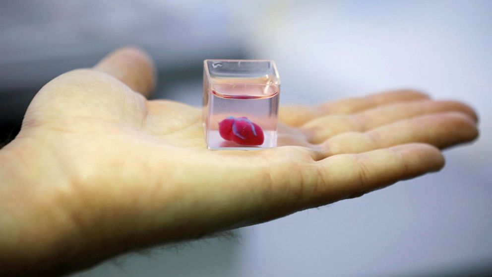
For the first time in history, scientists have created a three-dimensional, fully vascularized human heart. The biomedically engineered heart was created using a 3D printer by researchers at the University of Tel Aviv in Israel.
Modeled on a human patient, the 3D heart “[matches] the immunological, cellular, biochemical, and anatomical properties of the patient,” Dr. Tal Dvir, study researcher and professor of molecular cell biology at Tel Aviv University, said in a press release.
He added that the heart is made from human cells, and “patient-specific biological materials.”
“Our results demonstrate the potential of our approach for engineering personalized tissue and organ replacement in the future,” said Dr. Dvir.
Though it is still in the early stages of development, this invention represents a breakthrough for transplant medicine, as it may impact the lives of thousands of patients who await heart transplants for end-stage heart failure each year. A number of these patients will die while on the waiting list.
The engineered heart is about the size of a rabbit’s heart. As it continues to be redesigned to better reflect human anatomy, scientists are intrigued by the potential for 3D heart printing to become a widespread, life-saving technique in medical centers around the world.
This latest invention represents a major turning point for patients with congestive heart failure (CHF), as heart transplantation is the only definitive treatment for patients in the end-stages of the disease. CHF symptoms range from extreme shortness of breath to leg swelling and unintentional weight gain. These patients are at higher risk from sudden death relating to dangerous heart rhythms.
As such, CHF patients are frequently in-and-out of the hospital, require life-saving procedures to prevent dangerous heart rhythm, and suffer from a poor quality of life. Heart transplantation is oftentimes the only way to improve their quality of life and extend survival. Given the number of patients suffering from CHF each year, and its high healthcare costs, the study’s researchers were determined to “develop new approaches to regenerate the infarcted heart.”
The 3D heart was created from human cells obtained through biopsies. These tissue samples were experimentally reprogrammed to become “pluripotent” or de-identified stem cells. The stem cells were then exposed to chemicals or “bioinks” that helped to retrain them to become either heart or blood vessel cells.
“The biocompatibility of engineered materials [was] crucial to eliminate the risks of implant rejection, which jeopardizes the success of such treatments,” said Dr. Dvir.
Transplant rejection, which occurs when the recipient’s immune system targets transplanted tissue, is a common problem in heart transplant patients. It typically occurs within one year of heart transplantation, and accounts for a number heart transplant-related deaths.
Although the 3D human heart represents a promising step towards transplant engineering, further research is needed. The model needs to be studied in vivo, meaning in live organisms – through future animal studies – to understand its true biologic impact on the body, particularly in people with cardiovascular disease.
Navjot Kaur Sobti is an internal medicine resident physician at Dartmouth-Hitchcock-Medical Center/Dartmouth School of Medicine and a member of the ABC News Medical Unit.


